
Density of biological cells can be analyzed.
Neutrophil is known to undergo exocytosis of granul which has high index refraction at phagocytosis.
We visualize and analyze this granul distribution and exocytosis with qnatitative phase-brightfield illumination to the dual-imaging microscope.
Figure 7-1 indcates the configuration diagram of qunatitative phase-brightfield illumination to the dual-imaging microscope.
We introduced HOLORER-it (HL03G) as an addditinal illumination device for bright-field measurement and quantitative phase measurement at the same time.
Concentrating light HOLORER-it with the characteristics of high aperture ratio and long working distance can visualize high quality bright field image.
We can measure granule that has high index of refraction with quantitative phase measurement.
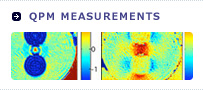
Figure 7-2 shows the time differentiation of the combined images of bright field image and quantitative phase image.
We can observe foreign substance in the neutrophil from bright field image.
As we observed with quantitative phase image, wide-range quantitative phase rate exists in the peripheral of the foreign substance. We can speculate that many granules exist around the foreign substance.
As we dynamically observed the combined images of bight field image and quantitative phase image, we can see the disappearance of granule around the foreign substance by exocytosis. And then we see the decrease of wide-range quantitative phase rate which was high on the whole.
Figure 7-3 shows the optical distance rate shift of granule when occurring exocytosis.
After exocytosis, optical distance rate decreased by about 40 nm in 20 mm second. And then we admitted the increase of optical distance rate.
In this way, we can quantitatively analyze very fast acting phenomena.
Quantitative phase microscope can non-invasively obtain quantitative data of living cells so that we can use this method for research and development tool on medical and development of new medicine field.
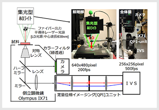
Figure 7-1. Dual-imaging of bright field and quantitative phase microscopy
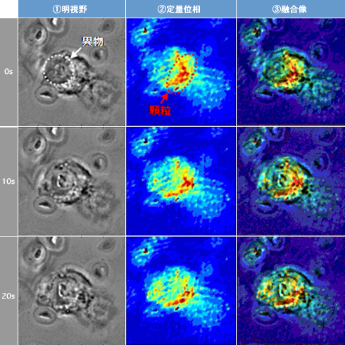
Figure 7-2. Merged images of bright field and quantitative phase
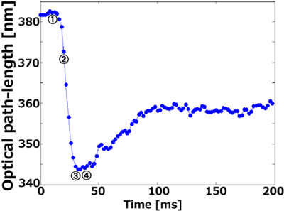
Figure 7-3. Optical length changing of granule during phagocytotic process in Neutorphil
Copyright © PiPhotonics, inc. All Rights Reserved.